A groundbreaking study published in Current Biology proposes a radical reinterpretation of vertebrate eye evolution, suggesting that our complex, paired eyes originated from a single, light-sensitive organ situated atop the head of an ancient ancestor. This research posits that the fundamental visual tissue predates the very existence of eyes as we know them, with evolutionary remnants still active within the human brain. This perspective challenges conventional understanding and offers a novel explanation for the unique visual systems found in vertebrates, setting them apart from the rest of the animal kingdom.
The fundamental distinction in light detection across the animal kingdom lies in two primary photoreceptor types: rhabdomeric and ciliary. Bilaterally symmetrical animals, characterized by distinct left and right halves, typically employ both. Rhabdomeric photoreceptors are commonly found in the paired eyes of invertebrates, facilitating image formation and navigation. In contrast, ciliary photoreceptors are often located deeper within the brain or as a singular dorsal eye, primarily regulating circadian rhythms and sensing ambient light levels. This dichotomy is observed in creatures ranging from insects and crustaceans to cephalopods, establishing a consistent evolutionary pattern for visual systems.
The Unique Vertebrate Visual System
Vertebrates, a diverse group including humans, birds, reptiles, and fish, exhibit a significant deviation from this established evolutionary blueprint. The vertebrate eye uniquely integrates both photoreceptor types. While ciliary cells are utilized for light capture within the vertebrate eye, the subsequent processing of these signals is handled by neurons possessing rhabdomeric characteristics. This hybrid cellular architecture is unprecedented in other animal lineages, leading to persistent questions within the scientific community regarding its evolutionary origins and functional integration.
The prevailing scientific consensus has long sought a cohesive explanation for the peculiar hybrid structure of vertebrate eyes. Questions have arisen about the ancestral state of vision and the extent to which subsequent species have modified or independently evolved their visual apparatus. The intricate neural pathways and cellular compositions within vertebrate retinas present a complex puzzle, prompting detailed investigation into the deep evolutionary history of sight.
Tracing the Origins: The Median Eye Hypothesis
To unravel this evolutionary enigma, researchers meticulously analyzed the placement and function of light-sensing cells across a broad spectrum of 36 major animal groups. Their findings suggest an evolutionary timeline tracing back to a worm-like ancestor that existed approximately 600 million years ago. This ancestral organism is hypothesized to have possessed both paired, lateral eyes positioned on the sides of its head and a singular, median eye situated on its dorsal surface.
The prevailing theory is that as these ancient creatures adopted a more sedentary, burrowing lifestyle to filter feed from the ocean floor, the selective pressure for complex, laterally placed eyes diminished. Maintaining these energy-intensive visual organs for navigation became biologically less advantageous. Consequently, the lateral eyes are thought to have gradually regressed and disappeared over evolutionary time. The single, median eye, however, remained essential for crucial functions such as regulating circadian rhythms and maintaining orientation, particularly in deep-water environments.
Repurposing and Migration: The Rise of Modern Eyes
The researchers propose that millions of years later, these organisms transitioned from their burrows back to open-water environments. This shift necessitated the re-establishment of sophisticated visual capabilities for navigation and predator detection. Given the absence of their original lateral eyes, evolution is thought to have ingeniously repurposed the remaining light-sensing structures. The ancestral median eye, situated on top of the head, gradually underwent a transformation.
This transformation involved the development of cup-like extensions capable of detecting the direction of incoming light. These structures are believed to have eventually bifurcated and migrated to the sides of the head, ultimately giving rise to the paired eyes characteristic of modern vertebrates. This evolutionary detour explains the unique cellular composition of vertebrate eyes, as the original median eye likely contained a mixed system of both ciliary and rhabdomeric cells. This hybrid circuitry was inherited by the newly formed lateral eyes, resulting in the multilayered retinas observed today.
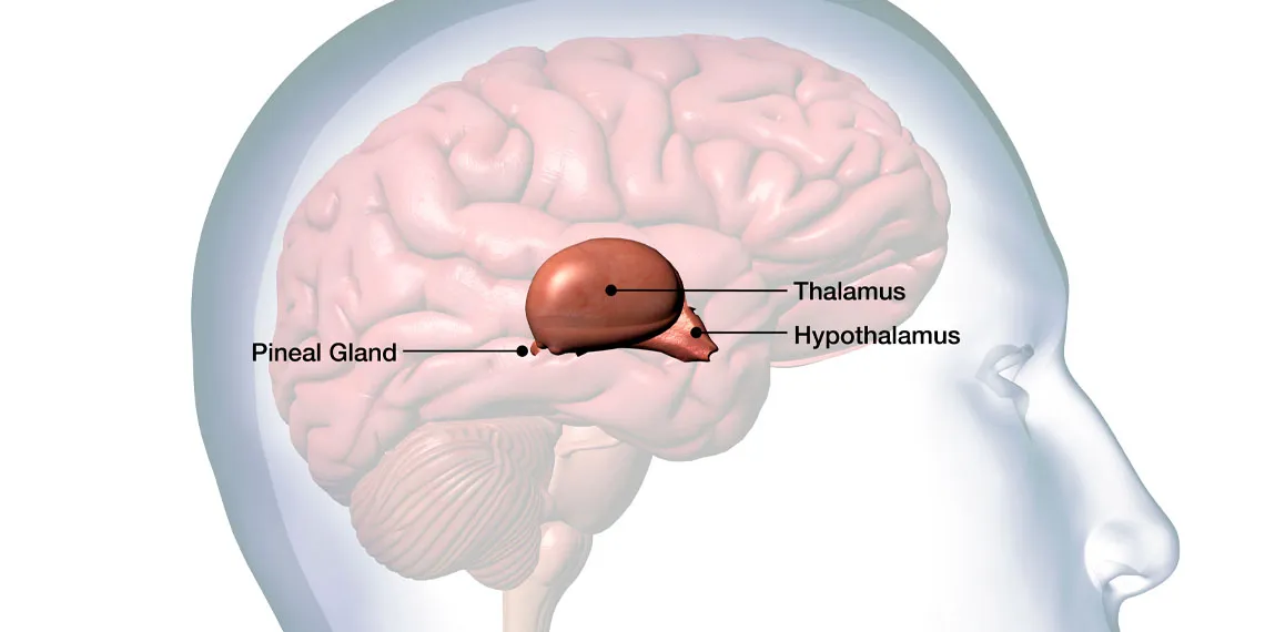
The Pineal Gland: A Living Relic
A critical component of this evolutionary narrative is the fate of the original median eye. The study suggests that this ancestral organ did not entirely vanish but rather persisted in an altered form. In mammals, this structure is believed to be represented by the pineal gland, a small endocrine gland located deep within the brain. While no longer directly processing visual input, the pineal gland continues to utilize light signals relayed from the eyes to regulate melatonin production and control circadian rhythms.
Evidence supporting this hypothesis can be observed in extant species. The tuatara, a reptile native to New Zealand, possesses a functional third eye on its crown, complete with a lens and retina, directly illustrating the ancestral median eye. In certain fish species, the pineal gland remains a more rudimentary organ capable of directly sensing light that penetrates the skull. This provides a tangible link to the evolutionary past, demonstrating the enduring legacy of the ancestral median eye.
Challenges and Future Directions
While this study presents a compelling and cohesive hypothesis, it is largely based on comparative analysis of cellular and genetic characteristics across extant species. The sparse fossil record from the period in question limits direct observation of the morphological changes that occurred in these ancient soft-bodied ancestors. Furthermore, the process of chimerization, where cellular traits have blended over millions of years, complicates the definitive tracing of precise evolutionary lineages for all neural circuits within the modern vertebrate retina.
Future research endeavors are expected to focus on expanding genetic datasets from a wider array of animal species to rigorously test these evolutionary propositions. Advanced imaging and mapping techniques will be employed to conduct detailed comparisons between the microscopic structures of the pineal gland and retinal tissues. This continued investigation aims to solidify or refine our understanding of the intricate evolutionary journey that shaped vertebrate vision.